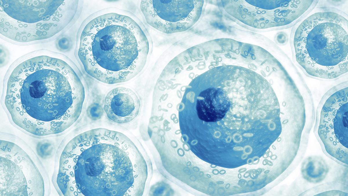
A landmark study by Indian researchers has not only found evidence of cells recovering from the near-death stage but has also found the molecular mechanism that drives the recovery process, the opposite of programmed cell death. Scientists at the Centre for Cellular and Molecular Biology (CSIR-CCMB), Hyderabad, call the survival mechanism that they uncovered as “Programmed Cell Revival”. “We here discovered a genetically encoded intrinsic revival code, which we named as Programmed Cell Revival, which allows cells to recover from a near-death state,” Dr. Santosh Chauhan, a senior researcher at CCMB and the corresponding author of a paper published in The EMBO Journal says.
Programmed cell death is generally considered as an irreversible process. However, in recent years, researchers have found evidence that some forms of cell death can indeed be reversed under certain conditions. The study by CCMB researchers led by Dr. Santosh Chauhan has for the first time uncovered the key molecular mechanisms that regulate the reversal of cell death.
Serendipity
The first signs of Programmed cell revival of cells were discovered by accident when Kautilya Kumar Jena, who was a PhD student about six years ago and now at BRIC-Institute of Life Sciences, Bhubaneswar, added a sublethal dose of a death-inducing agent (L-leucyl-L-leucine methyl ester — LLOMe) added to cause lysosome-mediated cell death in a petri dish, which led to revival of cells instead of causing death. The cells that are attached to the plate surface get detached and come into the surface and show cell death-like features. “The cells not only get detached but they also exhibit properties that suggest cell death has actually started, such as the bulging of the cell membranes (blebbing). The pathway to cell death appears committed. So, researchers collect the cells at this stage assuming that the cells are dead,” Dr. Chauhan. “The pathway to cell death is not limited to phenotypical features but molecularly also. We saw many molecular manifestations of cell death such as mitochondria getting disrupted or disintegrated.”
Though the cell death process had started but was not executed in full as only a sublethal dose of the death-inducing agent was used. Just five minutes after treatment with the death-inducing agent, the cells began the process of apoptosis as displayed by cells becoming round. Thirty minutes later, the cells began detaching from the growth surface and the cell membranes began bulging (blebbing), which is an advanced stage of cell death. Instead of further progressing towards programmed cell death, about 80-90% of the cells instead reattached to the surface within two-three hours, and regained their normal morphology in six hours. And 16 hours after treatment with the death-inducing agent, the cells appeared normal and began the division process. Remaining traces of abnormality disappeared in 24 hours, and the cells appeared morphologically normal.

As per literature, 4-8 millimolar of the death-inducing agent is considered sufficient to kill the cells. “But we found that at this concentration, the cell death process gets initiated but is not completely executed. The process of cell death then takes a U-turn and the cells don’t die but rebound when a sub-lethal concentration of the agent is used, which is really interesting,” Dr. Chauhan explains. The researchers found that cells can recover even when the integrity of the plasma membrane is compromised, but not after disruption of the nuclear membrane. They found that resuscitation from cell death was a highly regulated, well-programmed process.
Becoming stem-like cells
More than unravelling the reversal of cell death, the researchers discovered that the initiation of the programmed cell death actually causes the cells to reverse from a differentiated state to a de-differentiated state. “The cells appear de-differentiated, something like the embryonic stem cells. When the cells begin to die, they reprogramme themselves such that stem-like characteristics start appearing in them, and then they don’t die,” he says. “Since the cells don’t die, the reversal of cell death appears to be converting a fully differentiated cell into embryonic-like stem cells with a potential to regenerate and potential to heal.”
The reversal from differentiated state to a de-differentiated stem-like cells were not ascertained based on morphological characteristics alone but also molecularly. “We did whole genome RNA sequencing and epigenome studies to ascertain that the adult, differentiated cells were resetting themselves and becoming stem-like cells,” says Dr. Chauhan. “The data indicate that the reviving cells launch a molecular programme mimicking embryonic cells for reinitiating a new life,” they write.
Skin wound healing
Once the researchers observed that molecularly the cells appear stem-like, they proceeded to test the regenerative capacity of the cells that were treated using a sub-lethal dose of the cell death-inducing agent. First, the researchers tested the ability of the death-inducing agent (LLOMe) in speeding up skin wound healing in a mouse model.
The cell death-inducing agent was dissolved in water to get a concentration of 4 and 8 millioMolar, and the water was topically applied to the wound surface twice every day. The animals treated with LLOMe-containing water healed “significantly faster” compared with the control group. In just one day of treatment, the average reduction in wound size was 27% in the 4 milliMolar group and about 50% in the 8 milliMolar group, while the control group showed only 3% healing. By the third day, the average wound size was reduced by 64% and 78% in the 4 milliMolar and 8 milliMolar groups, respectively, compared with just 23% healing in the control group.
Corneal wound healing
Having tasted success with the mouse wound model, the researchers tested the effectiveness of the sub-lethal dose of the cell-killing agent in healing corneal injury. Collaborating with Dr. Kiran Kumar Bokara from CCBM who studies corneal injury models, the researchers tested the corneal wound healing in a mouse mode. The corneal epithelium of one eye was purposely damaged by gentle scraping of the corneal epithelium and alkali burn produced by topical application of a single drop sodium hydroxide. The cell death-inducing agent (LLOMe) was administered topically twice a day for seven days.

There was “significant reduction” of the epithelial defect from day two in the group treated with LLOMe compared with the control group. “We found that re-epithelisation, which is the kind of regeneration of the epithelial cells, was very fast and dramatically better in LLOMe-treated groups,” Dr. Chauhan says. The results indicate that the agent could prove to be a potent therapeutic option for treating corneal burn injuries.
Tadpole tail regeneration
“We were more encouraged by corneal wound healing results. Since both the skin wound healing and corneal wound healing are restricted to the surface, we wanted to test real regeneration potential,” he says. For this, they turned to the established tadpole tail regeneration model. In collaboration with Dr. Pravati Kumari Mahapatra from Utkal University, Bhubaneswar, the researchers tested the efficacy of LLOMe in accelerating a tadpole tail regeneration in Asian tree frog (Polypedates maculatus) model. The tails of Asian tree frog tadpoles were amputated and then treated using four different doses of LLOMe-containing water. They observed rapid blastema (cluster of undifferentiated cells that forms at the site of injury and has the ability to regenerate into an organ or appendage) formation in the treated group within 24 hours of amputation. The study found that treatment with different concentrations of LLOMe led to “significantly accelerated” tail regeneration compared with the control group. “In the case of tadpoles treated with sub-lethal concentration of the LLOMe agent, the tails were regenerating five-six days earlier than the control group,” Dr. Chauhan says. “We did some molecular experiments to understand what is happening in the tail during regeneration, and we found that the LLOMe agent was working through lysosomes.”
Axon regeneration
In one more test to verify the regenerative capacity of the sublethal dose of LLOMe agent, the researchers in collaboration with Dr. Anindya Ghosh-Roy of National Brain Research Centre (NBRC), Manesar, Haryana, tested the efficacy of the agent in inducing regeneration of axons in C. elegans. And they found that the regrowth length of axons after they were cut was “significantly enhanced” with LLOMe treatment. In addition, the functional restoration also improved “significantly” after the treatment.
Increase in haematopoietic stem cells
Finally, in collaboration with Dr. Rohan Jayant Khadilkar from the Advanced Centre for Treatment, Research and Education in Cancer (ACTREC), Tata Memorial Centre, Mumbai, the researchers tested the stem-like nature of cells when exposed to sublethal dose of LLOMe agent. The fruit fly larvae were treated with the cell death reagent at sublethal concentration (8 milliMolar) for 14 hours. The fruit flies showed “pronounced increase” in the haematopoietic stem and progenitor cells in the lymph glands upon LLOMe treatment.

“These new insights into the molecular mechanisms of programmed cell revival have promising therapeutic implications, while also opening new directions for experimental investigation,” says a News and View article in the same issue of the journal.
“The medical promise is immense, ranging from faster recovery after a stroke or heart attack to regenerating tissues in degenerative diseases. Instead of replacing damaged organs, doctors might one day coax the body to heal itself by awakening this hidden programme,” he says. “This breakthrough opens a new chapter in cell biology, blurring the line between death and repair and hinting at a future where healing comes from within. This study’s findings could be exploited in not just regenerative medicine and cancer, but also in ageing, neurodegeneration, and infection biology, to name a few
A word of caution
“But there’s also a warning. Cancer cells may exploit the same revival programme to resist treatment and return stronger,” he warns. Elaborating about the harm that such treatment may cause in people who have cancer he says: “The cancer therapies kill tumour cells. But chemotherapy or radiotherapy can be suboptimal leading to relapse of cancer. We feel relapse could be due to the procedure we discovered. In suboptimal dosing, cancer cells rather getting killed fully may revive more powerful with this programme and become stem-like, leading to therapy-resistant tumours. Ideally, drugs that can inhibit this process in combination with cancer therapies might be key to reduce cancer relapse,” he says.